| front |1 |2 |3 |4 |5 |6 |7 |8 |9 |10 |11 |12 |13 |14 |15 |16 |17 |18 |19 |20 |21 |22 |23 |24 |25 |26 |27 |28 |29 |30 |31 |32 |33 |34 |35 |36 |37 |38 |39 |40 |41 |42 |43 |44 |review |
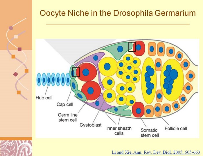 |
Li and Xie, Ann. Rev. Dev. Biol. 2005, 605-663
Drosophila germarium cross section showing the locations of germ line stem cells (GSCs), somatic stem cells (SSCs), and their niches. Two or three GSCs (red cells, left) are situated in their niche, composed of cap cells (green cells, left) and terminal filament cells (light blue cells, left tip), whereas their differentiated progeny, including cystoblasts and differentiated cysts (yellow cells, middle), are surrounded by inner sheath cells (purple cells and green cells, bottom and top). Two or three SSCs (red cells, bottom and top) directly contact the posterior group of inner sheath cells (green cells, bottom and top) forming their niche, whereas their differentiated progeny, also known as follicle progenitor cells (orange cells on right), further proliferate and generate differentiated follicle cells. Two inserts depict major signaling pathways controlling GSC (top and left) and SSC (top and right) self-renewal and proliferation; these inserts also depict niche cells (green) and stem cells (pink). |