| front |1 |2 |3 |4 |5 |6 |7 |8 |9 |10 |11 |12 |13 |14 |15 |16 |17 |18 |19 |20 |21 |22 |23 |24 |25 |26 |27 |28 |29 |30 |31 |32 |33 |34 |35 |36 |37 |38 |39 |40 |41 |42 |43 |44 |review |
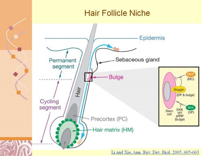 |
Li and Xie, Ann. Rev. Dev. Biol. 2005, 605-663
Illustration of the epidermal stem cells. Stem cells are located in the bulge region of the hair follicle beneath the sebaceous gland. Upon activation, stem cells undergo division; the daughter cells retained in the bulge remain as stem cells while other daughter cells migrate down to become hair-matrix progenitors responsible for hair regeneration. In neonatal mice or in damaged skin, stem cells can also migrate upward and convert to epidermal progenitors that replenish lost or damaged epidermis. The bulge area is an environment that restricts cell growth and differentiation by expressing Wnt inhibitors, including DKK,Wif, and sFRP as well as BMPs. During the early anagen phase, Wnts from dermal papilla (DP) and Noggin, which is derived from both DP and bulge (J. Zhang & L. Li, unpublished data), coordinate to overcome the restriction signals imposed by both BMPs and Wnt inhibitors; this leads to stem cell activation and subsequent hair regeneration. The FGF and Notch pathways are also involved in DP function for hair-matrix cell proliferation and lineage fate determination, but their influence on stem cells is not clear. |