
Subscribe to Pittwire Today
Get the most interesting and important stories from the University of Pittsburgh.Childbirth is a momentous and fraught time. It’s also one of the most significant biomechanical events in life. In terms of forces and motion, the birthing body is a structure stressed to extremes.
For bioengineer Steven Abramowitch, that is not a cold description. He studies damage to people’s pelvises due to pregnancy and delivery — damage that may only manifest decades after the birth. His team creates computer models of pelvic structures that could indicate potential health problems.
Abramowitch says women’s health research has too often played second fiddle or been considered too private to discuss: “Pelvic prolapse — when pelvic muscles can no longer hold up organs like the bladder — is more prevalent than conditions like ACL injuries, but no one discusses it. It’s embarrassing. It can lead to symptoms like urinary and fecal incontinence, which folks don’t like talking about.”
Abramowitch is associate professor of bioengineering at the Swanson School of Engineering and the Clinical and Translational Science Institute. His research into pelvic health disorders increasingly relies on computer modeling, which beats animal models for a simple reason — the animal models are quadrupeds. Gravity does not affect their organs the ways it affects humans. Bipedal humans inherited an evolutionary conflict between efficiently walking upright and efficiently delivering babies.
“Bodies in childbirth are really at the limits of what the tissues can withstand,” Abramowitch explains. “Childbirth is associated with a lot of soft tissue injuries, many of them unrecognized because they are internal. A muscle tear or nerve damage is not obvious to the obstetrician delivering the baby.”
The injuries appear later in life, sometimes driven by a secondary event or in a slow degenerative process. Modeling similarities and differences among a range of pelvic shapes, Abramowitch believes, can provide insight into the potential for injury.
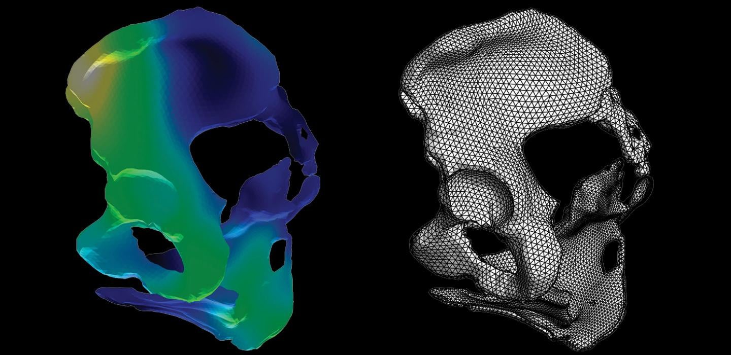
Abramowitch’s team uses several computational steps to model the pelvis and bones of the hip, sacrum and coccyx, based on MRI scans. They essentially break the complex whole of the scan into many small geometric points — more points create a more complete picture — then build a statistical model of the points distribution. The shapes that have been broken into small pieces are reassembled and compared.
“With the models we can say what anatomical features are associated with certain types of birth outcomes,” explains Abramowitch. “We try to find underlying mechanisms.”
PhD candidate Megan Routzong is part of Abramowitch’s team. “When I started this work, some family asked: ‘Why are you studying childbirth? Women have been giving birth for centuries,’ I ask, ‘Did they enjoy that experience? How many women still die from that experience or have a severe quality-of-life loss?’”
“The idea is that if we define an average or typical anatomy, the more easily a clinician would be able to identify anatomy that tends toward a disease state. The question is which anatomical factors — computationally we refer to them as geometries — correspond with predicted negative vaginal birth outcomes, like excessive stretching in one region of a muscle.”
People typically get ultrasounds during pregnancy. With this model it would be possible to use the portion of a pelvis visible in the ultrasound field of view — smaller than an MRI field of view — to make predictions about how the entire pelvic shape might affect the delivery. The goal is reducing the amount of data needed to make predictions, ultimately to create a routine clinical tool.
For Routzong, the free advanced computing resources available to Pitt researchers were vital to the project. “This research would not be feasible without those resources. We didn’t need to already be funded to create a preliminary study. We did the modeling at the Center for Research Computing on a scale that not possible on any of our machines.”
Abramowitch considers the potential impact of the work.
“I’m a trained bioengineer, so these problems are very interesting to me from that perspective. But they are quality-of-life issues. Women are deeply affected. The long-term impact of pelvic damage is not just a casual part of the aging process, like wrinkles or walking slower. These can be really catastrophic, life-altering conditions.”
This story was written by Brian Connelly in the Center for Research Computing.