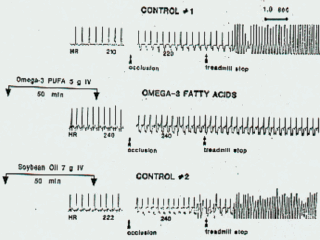
Figure 2. Representative ventricular electrogram from the same dog with and
without intravenous infusion of n-3 fatty acids. Control 1:
exercise-plus-ischemia test done 1 week before an exercise-plus-ischemia
test immediately preceded by i.v. n-3 fatty acids. Control 2 repeated 1 week
after infusion of n-3 fish oil fatty acids. This time a lipid emulsion
derived from soybean oil (Intralipid) lacking free n-3 fatty acids was
infused. |
Figure 2 illustrates the typical response of
one of the susceptible dogs to the exercise-ischemia protocol. The top
control No.1 tracing is an electroventriculogram. Because the dog was
running on the treadmill, its pulse rate is elevated. The additional
ischemic stress from occlusion of the left circumflex artery for 2 minutes
resulted in a ventricular tachyarrhytmia and the circulation failed. As soon
as the dog lost consciousness the dog was defibrillated. The second tracing
is of the same dog brought back into the laboratory one week later, when the
same protocol was repeated. This time just before the left coronary artery
was occluded a phospholipid emulsion containing free n-3 fatty acids was
infused intravenously. Additional ischemic stress from occluding the left
circumflex coronary artery this time failed to induce VF. One week later,
control No. 2 was performed on the same dog. An emulsion of soybean oil,
which lacks any free n-3 fatty acids, was infused. Within 2 min of occluding
the left circumflex artery, VF occurred. This is the protocol we used to
test the antiarrhythmic effect of the n-3 fish oil fatty acids with a
control one week before and one week after the test with the infusion
intravenously of the fish-oil-free fatty acids. |