| front |1 |2 |3 |4 |5 |6 |7 |8 |9 |10 |11 |12 |13 |14 |15 |16 |17 |18 |19 |20 |21 |review |
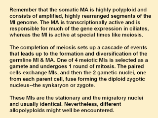 |
After formation of the zygote, the MI undergoes
2 rounds of mitosis, producing 4 new diploid nuclei per cell. However this
behavior varies a great deal among the species of ciliates. Example: first
zygotic division (FZD) could degrade 1/2 MIs and proceed with one. At this
stage at 8 h in conjugation, there are 4 zygotic nuclei in Tetrahymena. The old parental MA moves to the bottom of the cell and eventually goes through apoptotic degradation. Still, it could be recalled to control the cell if the new MA failed. Of the 4 nuclei, 2 become somatic MA, whereas the other 2 remain germline MI very soon after this decision point, the CNA1p localization in the 2 nuclei destined to become MIs reverts to the localization pattern seen in interphase MI. In these 2 nuclei, CNA1p is seen in fewer than 10 dots. P stands for protein. In the nuclei destined to become MA, CNA1p decreases as points throughout the nucleus, as conjugation progresses until genomic rearrangement when the staining disappears entirely in the developing MA. Meanwhile, bright staining of CNA1p is seen late in the apoptotic degradation of the old MA, when most DNA has been degraded. Intense CNA1p staining corresponds to CNA1p that moves from the developing MA to the old parental MA to be degraded. Pdd1p that is involved in DNA elimination in the developing MA, is similarly localized to the parental MA before being degraded This early differentiation included the removal of the MI-specific linker histone, the appearance of a MA histone H2A variant and acetylation of histone H4 in those nuclei destined to become MAs. |