| front |1 |2 |3 |4 |5 |6 |7 |8 |9 |10 |11 |12 |13 |14 |15 |16 |17 |18 |19 |20 |21 |22 |23 |24 |25 |review |
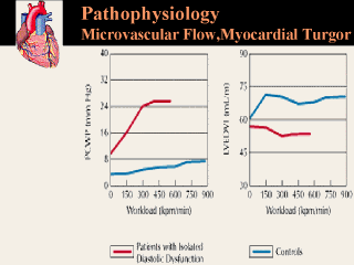 |
Figure 3. In studies of patients with isolated diastolic dysfunction pulmonary capilary wedge pressure (PCWP) was elevated at rest and further elevated during exercise (left). Although this indicates that left ventricular filling pressure was also increased under both conditions, that was not reflected in the left ventricular end diastolic volume index (LVEDVI), which failed to increase with exercise (right). (Adapted from Kitzman et al, 1991) |