| front |1 |2 |3 |4 |5 |6 |7 |8 |9 |10 |11 |12 |13 |14 |15 |16 |17 |18 |19 |review |
 |
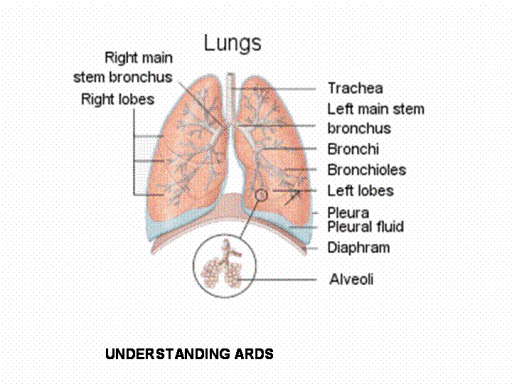
To understand ARDS, it is important to review how the
lungs work. Air, which contains oxygen, is inhaled through the nose
and mouth, and passes into the windpipe (trachea). From the trachea,
air flows through tubes called bronchi into microscopic air sacs
called alveoli. Very small blood vessels (capillaries) are imbedded
in the walls of these air sacs. Oxygen passes through the thin walls
of the alveoli into the bloodstream. Carbon dioxide, a waste product
of cellular function throughout the body, passes from the
bloodstream into the alveoli and then is exhaled. At the onset of
ARDS, lung injury may first appear in one lung, but then quickly
spreads to affect most of both lungs.
When alveoli are damaged, some collapse and lose their
ability to receive oxygen. With some alveoli collapsed and others
filled by fluid, it becomes difficult for the lungs to absorb oxygen
and get rid of carbon dioxide.
Within one or two days, progressive interference with gas
exchange can bring about respiratory failure requiring mechanical
ventilation.
As the injury continues over the next several days, the
lungs, fill with inflammatory cells derived from circulating blood
and with regenerating lung tissue.
Fibrosis (formation of scar tissue) begins after about 10
days and cam become quite extensive by the third week after onset of
injury. Excessive fibrosis
further interferes with the exchange of oxygen and carbon dioxide.
The sequential stages of ARDS are described in further detail
below.
|