| front |1 |2 |3 |4 |5 |6 |7 |8 |9 |10 |11 |12 |13 |14 |15 |16 |17 |18 |19 |20 |21 |22 |23 |24 |review |
 |
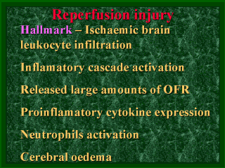
The reperfusion injury that
follows the biochemical cascade is driven by cytokines and leukocytes. The hallmark of an
inflammatory reaction in the ischaemic brain is leukocyte infiltration, which is dominated
by polymorphonuclear leukocytes (PMN), primarily neutrophils (Feuerstein et al.
1994). Ischaemic vessels in the brain are filled with leukocytes and many have oedema
around them. The adhesion of leukocytes to the walls of cerebral blood vessels and their
infiltration into ischaemic brain tissue activates an inflammatory reaction, which results
in the release of large amounts of oxygen-free radicals, including superoxide anions,
hydrogen peroxide, hydroxyl radicals and singlet oxygen. This reaction exacerbates the
degree of tissue injury. Leukocytes also interfere with normal microvascular perfusion.
(Feuerstein et al. 1998) Cytokines are a large and rapidly expanding group of polypeptides produced by many cell types. They are mediators of immuno-endocrine interactions and are necessary for the optimal functioning of leukocytes. The cytokines are key mediators of acute phase responses to tissue injury (Tonnesen et al. 1996), and the pro-inflammatory cytokines induce increased neutrophil and endothelial surface adhesive molecule expression, promoting neutrophil-endothelial adherence. This adherence and subsequent neutrophil organ binding is thought to be a “common pathway” for organ injury (Hill et al. 1997). An important source of cytokines in the central nervous system (CNS) is the peripheral blood cells (e.g leukocytes) which may enter the brain after injury and ischaemia. Cytokines are also produced by resident brain cells, including glia, neurones and endothelial cells (Rothwell and Strijbos 1995). Interleukin 1 (IL-1) has been shown to mediate neurodegeneration in vivo (Rothwell and Strijbos 1995), and tumour necrosis factor alpha (TNF- a) and IL-1betta seem to play a significant role in brain immune and inflammatory activities and ischaemic brain injury (Feuerstein et al. 1998). Activated neutrophils may induce multi-organ oedema, including cerebral oedema, via TNF-a and other regional cytokines. (Dewanjee et al. 1998)The presence of recruited leukocytes at the site of inflammation is critically dependent on co-ordinated expression of the adhesion molecules. The efficient “docking “ of activated leukocytes to their respective receptors on inflammatory cells and the activated capillary is the primary step in the process of transendothelial migration. The candidate molecules include ICAM-1, ELAM-1 and P-selectin on the endothelial side and CD11/CD18, MAC-1 and LFA-1 on the leukocyte side. (Feuerstein et al. 1998) |
| front |1 |2 |3 |4 |5 |6 |7 |8 |9 |10 |11 |12 |13 |14 |15 |16 |17 |18 |19 |20 |21 |22 |23 |24 |review |