| front |1 |2 |3 |4 |5 |6 |7 |8 |9 |10 |11 |12 |13 |14 |15 |16 |17 |18 |19 |20 |21 |22 |23 |review |
 |
Most of
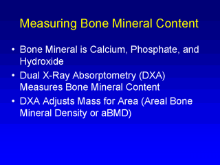
bone mineral is hydroxyapatite: calcium, phosphate, and hydroxide. Calcium is
approximately 32% of bone mineral mass. Though bone mass is more than just minerals,
dual-energy x-ray absorptiometry (DXA), the leading technology used in bone-related
research, captures only bone mineral and not true bone mass. Using DXA , various regions of the skeleton can be assessed, although most often the lumbar spine, the proximal head of the femur, or the total body (entire skeleton) are used in research and clinical assessments. The quantitative measure of bone mass (bone mineral content) provided by the DXA scan is a function of the size of the bone as well as of the amount of mineral within the bone. Therefore, bone mass (in grams) is adjusted for bone size by dividing by bone area (cm2, i.e., length and width). This parameter is termed areal bone mineral density (aBMD) and provides a partial adjustment for bone size. In adults, fracture rates are highly associated with low aBMD measurements. |