| front |1 |2 |3 |4 |5 |6 |7 |8 |9 |10 |11 |12 |13 |14 |15 |16 |17 |18 |19 |20 |21 |22 |23 |24 |25 |26 |27 |28 |29 |30 |31 |32 |33 |34 |35 |36 |37 |38 |39 |40 |41 |42 |43 |review |
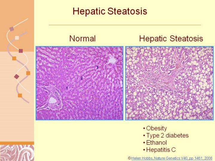 |
© Helen Hobbs, Nature Genetics V40, pp 1461, 2008
Study in this area is hindered by the lack of a clear consensus regarding the nomenclature and histologic categorization of NAFLD and NASH. However, most experts in the field do agree that in addition to steatosis and inflammation, ballooning degeneration and fibrosis are required features of NASH. The liver histology of NASH is characterized by the following 1) Macrovesicular fat deposits. Cytoplasmic lipid droplets composed principally of triglycerides and some cholesteryl esters that stain positively with Oil red-O stain. 2) Ballooning degeneration. Hepatocellular injury results in two different morphologic manifestations, either ballooning degeneration or acidophilic degeneration. The ballooning results from intracellular fluid accumulation and the cells are typically located in zone 3 (pericentral). 3) Evidence of focal necrosis with mixed polymorphonuclear inflammatory cells. The inflammation of NASH is typically mild and is predominantly lobular rather than portal. Neutrophilic cells in the lobular inflammatory infiltrates are a distinguishing feature from other forms of acute and chronic liver injury Sinusoidal fibrosis. The patterns of fibrosis are one of the characteristic findings in NASH. The deposition of collagen initially occurs in the perivenular and perisinusoidal spaces of zone 3. In some areas the collagen envelopes single cells in a pattern that is commonly referred to as chicken-wire fibrosis. This pattern of fibrosis distinguishes NASH and alcohol-induced fibrosis from other forms of chronic liver diseases in which the fibrosis is initially periportal. 5) Mallory bodies. Mallory’s hyaline is an intracytoplasmic inclusion that consists of many aggregated cytoskeletal peptides, some of which include cytokeratins 7, 18, 19 and ubiquitin. It is generally located in ballooned hepatocytes in zone 3. If found in setting of #’s 1-4, it is very supportive, but not required, for the diagnosis of NASH. In adult studies, the incidence of Mallory’s hyaline ranges from 9.5-90% (10, 11).
|