| front |1 |2 |3 |4 |5 |6 |7 |8 |9 |10 |11 |12 |13 |14 |15 |16 |17 |18 |19 |20 |21 |22 |23 |24 |25 |26 |27 |28 |29 |30 |31 |32 |review |
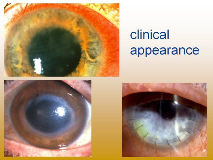 |
Upper photo shows stromal infiltate in upper 2/3rds of cornea and adjacent scleral inflammation Lower left photo shows classic ring infiltrate but no scleral involvement Lower right photo shows post-op corneal transplant, enlarged nearly to sclera in effort to capture entire infection. Sutures and incision near sclera and its vascular supply greatly increase risk of transplant rejection. |