| front |1 |2 |3 |4 |5 |6 |7 |8 |9 |10 |11 |12 |13 |14 |15 |16 |17 |18 |19 |20 |21 |22 |23 |24 |25 |26 |27 |28 |29 |30 |review |
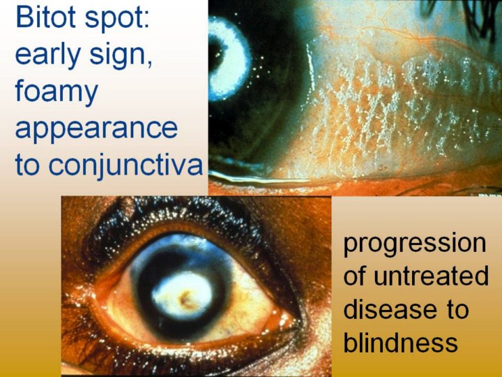 |
Upper photos: Bitot spot on conjunctiva, nasal and temporal exposed areas. Also note poor photo flash light reflection on right photo, due to poor tear film. Lower series of photos show (clockwise from upper left): Bitot spot; exposure ulcer (with fluorescein stain); larger sterile ulcer from poor tear film and chronic dryness; end-stage central scar from healed infection |