 |
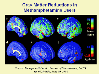
Figure 2. Gray-matter differences on the
lateral brain surfaces. The mean reduction in gray matter in the MA group,
relative to healthy controls, is expressed as a percentage and shown
color-coded (blue colors, no reduction; red colors, greater reduction). In
the left medial wall (a) and right-lateral (b) and left-lateral (c) brain
surfaces, gray-matter differences are not pronounced. The significance of
these differences is plotted in d-f. Differences were not significant after
correction for multiple comparisons. |
