 |
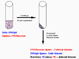
Figure 8. Determining intravesicular (or
intracellular) space. A concentrated suspension of vesicles or cells of a
known protein concentration is mixed with ³H₂O
or [³H]urea which distribute equally
between the internal and external spaces and [14C]sucrose which is
impermeant. The vesicles or cells are then centrifuged, the supernatant is
aspirated, the walls of the tube are dried, the pellet is resuspended and an
aliquot is assayed for ³H and 14C in a
scintillation counter. The difference between the
³H space (i.e. the internal plus
external spaces) and the [14C]sucrose space (the external space) is the
internal volume.
Knowing the protein concentration, the internal volume can be expressed in
µL per mg protein. From this value,
substrates taken up by the vesicles or cells can readily be converted to an
internal concentration. Since the external concentration is known (the
amount added minus the amount taken up), the concentration gradient can be
calculated. |