 |
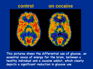
Positron Emission Tomography (PET) Scan of a
Person Using Cocaine
Cocaine has other actions in the brain in addition to activating the brain’s
reward circuitry. Using brain imaging technologies, such as PET scans,
scientists can see how cocaine actually affects brain function in people.
PET allows scientists to see which areas of the brain are more or less
active by measuring the amount of glucose that is used by different brain
regions. Glucose is the main energy source for the brain. When brain regions
are more active, they will use more glucose and whey they are less active
they will use less. The amount of glucose that is used by the brain can be
measured with PET scans. The left scan is taken from a normal, awake person.
The red color shows the highest level of glucose utilization (yellow
represents less utilization and blue indicates the least). The right scan is
taken from someone who is on cocaine. The loss of red areas in the right
scan compared to the left (normal) scan indicates that the brain is using
less glucose and therefore is less active. This reduction in activity
results in disruption of many brain functions. |