 |
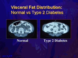
As seen in computed
tomographic (CT) scans, the distribution of visceral fat (white areas)
differs in a subject with normal glucose tolerance (left) and in
a subject with type 2 diabetes (right). The two subjects had similar waist
circumferences, but the individual with type 2 diabetes had a larger amount
of visceral fat than did the subject with normal glucose tolerance. The
amount of subcutaneous fat was larger in the subject with normal glucose
tolerance than in the subject with type 2 diabetes.
Go to Part II of this lecture
 |
Go to Comment Form |
|