| front |1 |2 |3 |4 |5 |6 |7 |8 |9 |10 |11 |12 |13 |14 |15 |16 |17 |18 |19 |20 |21 |22 |23 |review |
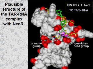 |
Specifically, the
arginine on ring I conjugated via the aliphatic amine of neomycin B is
positioned in the cavity formed by the bulge of TAR-RNA.
The surface of TAR-RNA is colored WHITE. The phosphate groups are RED. The neomycin core of NeoR is shown in GREEN.The arginine moieties are in YELLOW. The guanidino group of the arginine on ring I interacts with atoms N7 and O6 of guanine 26, whose surface is shown in MAGENTA.The a-amino group interacts with the O2P atoms of guanines 21 and 36 of TAR-RNA. The structure of the TAR-RNA is based on the structure of the TAR-RNA-arginine complex (PDB code 1arj). The right panel is a ~90 degrees turn clockwise of the left panel. |