
The Ligament Lengths of the Bundles of the ACL: A Computational Approach
Maribeth Thomas, Eric K. Wong, BS, Richard E. Debski, PhD
Musculoskeletal Research Center
Department of Orthopaedic Surgery
University of Pittsburgh Medical Center, PA
Introduction
The stability of the knee is due to its associated ligaments and muscles. The anterior cruciate ligament (ACL) has been shown to be the primary stabilizer for anterior tibial translation. It is also a secondary stabilizer for internal and valgus rotations and consists of two functional bundlesčthe anteriomedial (AM) bundle and the posteriolateral (PL) bundle.
The ACL is the most frequently torn ligament of the knee. ACL injuries most commonly occur while playing sports such as basketball, tennis, skiing, and football. Torn ACL ligaments are usually repaired through reconstructive surgery.
To investigate the results of reconstructive surgery, computational models can be used to simulate joint motion and to predict the ligament elongation under specific loading conditions. This can then be used to determine in-situ forces when combined with tensile testing data. The goal of this project is to develop a computational model to determine the ligament lengths of the bundles of the ACL during various joint motions.
Methods and Materials
To construct the computational model of the knee, a cadaveric specimen was used to obtain the bone geometry, kinematic motions, and ligament insertion sites as shown in Figure 1.
Experimental Load-Elongation Data
Figure 1. A Flow Chart of the Input and Resulting Output of the Computational Model of the Knee and ACL Ligament [1]
Two reference blocks were used to establish a common coordinate system. Two individual sets of data were collected for each reference blockčthe data obtained from the CT Scan and the data obtained from digitizing points on three orthogonal faces with the MicroScribe-3DTM (Immersion Corporation, San Jose, CA) tool. The Microscribe was also used to digitize the insertion sites of the ligament. The blocks were used to establish a transformation between the CT and Microscribe coordinate system. Four Mathematica programs converted the CT Scan reference block data points into the Microscribe-reference block coordinate system. Once the transformation was created, both the femur and tibia bone files were converted to that coordinate system.
After the coordinates of the reference blocks were established, the bone files were converted to a format readable by SIMM (Software for Interactive Musculoskeletal Modeling, Musculographics, Inc, Evanston, IL) using Microsoft Excel [3]. Finally, the file was run through SIMMšs Norm program that checked the data lists for inconsistencies and filled in any possible holes in the bone surface.
Four types of files made up the SIMM computational modelča bone file, a joint file, a motion file, and a muscle file. The bone file, as previously described, contains the vertices of the triangular polygons and the connections of the bone. The motion file provided the model with kinematics by defining the movement of the joint and the range of motion under different loading conditions from a previous robotic experiment. The muscle file defined the coordinates of the ligament insertion and created a simulated ACL ligament. The joint file defined the coordinate system, the axes of motion, and the Euler angle sequence for rotation.
Our computational included the femur, tibia, and a simulated ACL with two bundles. The kinematics were then simulated with the model, including anterior tibial load (simulated Lachman tests) at 15░ knee flexion and combined valgus and internal rotation (simulated pivot shift test) at 15░. ACL bundle length was measured under the testing conditions, and results were plotted in SIMM using the Plot Maker tool. Resulting data was plotted to compare bundle lengths.
Results
Figure 2 shows a picture of the completed model.
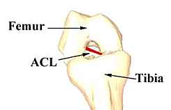
Figure 2. The SIMM Musculographic Model of the Knee and the ACL bundles
For the simulated Lachman test, the AM and PL varied at different knee angles of flexion and extension. In each case, the length of the ligament never exceeded 30 mm.
At 0░ of knee flexion, both ACL bundles showed similar trends as the length of both bundles increased with an increasing load. The AM bundle has a longer length at the reference position, and both the AM and PL bundles showed little change in length as the force increased to full anterior load.
For the pivot shift test, the knee was first positioned at 15 degrees of knee flexion. The AM and PL bundles showed maximum length at full internal rotation and minimum length at the 0 position.
Figure 3. Bundle Length vs. % of 134 N Anterior Load for a simulated Lachman test at 15 deg. of knee flexion
Discussion
The results from both the simulated Lachman test and the pivot shift tests show that the PL bundle does lengthen when an external load is placed on the knee joint. Although it appears that the AM bundle is longer, it also has a longer reference length since it may actually have a higher force. Further tensile testing is needed to compare in-situ forces in the bundle. Once validated, additional loading conditions can be attempted. However, from this data, it does appear that both bundles are lengthening and taking up load.
References
1. Debski, RE, J. Biomech Eng. 121(3):1999, 311-5.
2. Pfaeffle, HJ, J. Biomech. 32(12):1999, 1331-5
3. Vangura, AL- M.S. Thesis, Univ. of Pitt., 2000
Acknowledgments
I would like to thank the following people for making my summer research experience at the MSRC possible: Eric Wong, Dr. Savio L.Y. Woo, Dr. Richard Debski, Dr. Lars Gilbertson, Mary Gabriel, Kitty Stabile, My Fellow Summer Students.