
Development of a New Laser Micrometer System to Measure the Cross-Sectional Area of Tissue
Kristina Goodoff, Rich Debski, Ph.D.Musculoskeletal Research Center
Department of Orthopaedic Surgery
University of Pittsburgh Medical Center, PA
Introduction
A reliable technique for measuring cross-sectional area of a tissue is important in determining the stress a tissue can withstand. Change in cross-sectional area of the healing ligament must also be measured accurately in ligament healing studies. In a system used previously, the specimen rotated in the laser beam. This motion of the specimen caused inaccurate readings due to vibration of the specimen. The old system also had slow measurement and reconstruction processes as well as unreliable communication with the laser. Therefore a system was developed in which the cross-sectional area of a tissue could be accurately determined by placing the tissue in the path of a collimated laser beam and taking readings at certain increments as the laser is rotated around the tissue using a stepper motor (Stepper Motor HT23-401, Applied Motion Products, Inc.). The design objectives of the new system included (1)improving the speed of the measurement and reconstruction processes by using a faster laser and computer, (2)creating a reliable and user-friendly program to take measurements, and (3)allowing the specimen to remain undisturbed as readings are taken.
Methods and Materials
A program was developed using Microsoft Visual Basic 5.0, which allows the user to send commands to the motor driver (Panther LE2, Intelligent Motion Systems, Inc.). The driver was sent commands from the computer in the form of ASCII characters and numerical values designated by the user. Small programs were written into the driverís non-volatile memory that consisted of a series of commands to be executed by a single command issued by the user.
A separate program was then written to collect data from the laser (LS-3060, Keyence Corp.) using the shared memory file ShareData.dll provided by TLAser, Inc. The laser collects data at a rate of 400 scans/sec. Any number of scans specified by the user can be averaged to obtain one reading of the profile width and a reference distance. These readings are then processed to obtain one measurement of the cross-sectional area of an object.
The laser system collected data using the shadow method in which the specimen is placed in a collimated laser beam (see Figure 1). The diameter or profile width of the object as well as a reference distance was collected by the laser.
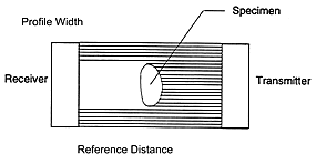
Figure 1: Laser heads (transmitter and receiver) and shadow (profile width) created by specimen in the collimated laser beam
The accuracy and repeatability of the system were then tested using three geometric shapes: triangle, square, circle. To determine the appropriate number of scans to average, static scans were taken of a circle at 1, 10, 20, 50, and 100 scans averaged. For the static scans, the placement of the circle in the laser beam was held constant as the number of scans averaged was varied. The laser was not rotated around the object during these readings so that only the most accurate setting for the laser could be determined. The cross-sectional area of each shape was then measured with the laser averaging 10 scans and then 50 scans while maintaining a constant orientation with the shape for 10 measurements. The areas of the square and triangle were measured again at 10 averaged scans and 50 averaged scans, however the orientation of the object within the collimated laser beam was changed for each of the 10 measurements. The same measurements were taken for the circle, but instead of changing the orientation, the placement of the object in the beam was varied. Measurements were also taken using a transparent cylinder in which the transparency of the cylinder was varied for several trials. Ten measurements were taken of the completely transparent cylinder. The cylinder was then scratched using sand paper and ten more measurements were taken. This process was repeated twice until the cylinder had been scratched three times. The degree to which the cylinder was scratched was measured on a scale ranging from 0-3.
Results
Based on the standard deviations of the reference distances, averaging 50 scans was found to give the most accurate area measurements of the circle when the static scans were taken. At one scan averaged, 20 scans averaged, and 50 scans averaged, the standard deviations of the reference distances were .015, .002, and .001 respectively. The measurements taken of the geometric shapes are listed in Tables 1-3. The data collected on the transparent cylinder are plotted in Figure 2. For the testing of the geometric shapes, the area measurements of the triangle were the least accurate. The measured area of the square was reasonably accurate and the area of the circle was most accurate. The most accurate areas for the triangle, square, and circle were taken at 50 averaged scans and variable orientation, 50 averaged scans and constant orientation, and 50 averaged scans and variable orientation, respectively. For the transparent cylinder, the most accurate area was achieved after the cylinder was scratched only once, however, there was little change in the error after the cylinder was scratched two and three times.
|
Triangle
|
Area(mm) | %Error | ||
| Orientation | Constant t | 10 Scans Ave. | 241.25 Ý 3.61 | 8.10 |
| 50 Scans Ave. | 240.08 Ý 2.63 | 7.58 | ||
| Variable | 10 Scans Ave. | 236.91 Ý 3.43 | 6.16 | |
| 50 Scans Ave. | 235.62 Ý 4.01 | 5.58 | ||
Table1: Cross-sectional area measurements of triangle
|
Square
|
Area(mm) | %Error | ||
| Orientation | Constant t | 10 Scans Ave. | 166.82 Ý .39 | 1.91 |
| 50 Scans Ave. | 165.17 Ý 1.22 | 0.90 | ||
| Variable | 10 Scans Ave. | 165.26 Ý 1.55 | 1.00 | |
| 50 Scans Ave. | 166.02 Ý 1.20 | 1.42 | ||
Table 2: Cross-sectional area measurements of square
|
Circle
|
Area(mm) | %Error | ||
| Orientation | Constant t | 10 Scans Ave. | 127.20 Ý .09 | 0.11 |
| 50 Scans Ave. | 127.24 Ý .14 | 0.08 | ||
| Variable | 10 Scans Ave. | 127.26 Ý .10 | 0.06 | |
| 50 Scans Ave. | 127.32 Ý .03 | 0.02 | ||
Table 3: Cross-sectional area measurements of circle
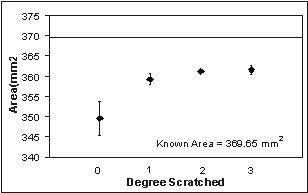
Figure 2: Cross-sectional area measurements of transparent cylinder
Discussion
A system to take accurate cross-sectional area measurements of a specimen was successfully developed. The areas of the triangle and square were overestimated due to the method used to calculate the area. When the area is calculated the maximum profile width is always used causing the area to always be overestimated2 even though the reconstructed image appears to have a smaller area due to rounding of the corners as shown in Figure 3. Therefore, an accurate image reconstruction of an object with sharp corners is dependent on the orientation of the object in the laser beam. Certain orientations produced images with more rounded corners while others produced images with sharp corners. However, a highly accurate area is measured for more rounded objects without sharp edges. A major limitation of the shadow method is that concavities are not detected during the reconstruction of the area because a collimated laser beam is unable to pick up concavities present in an object. It was shown that the area of semi-transparent objects can also be accurately measured using the laser. However, an accurate area measurement could not be taken of a completely transparent object. Further testing on biological tissue must still be done using the new system. Also, a clamping system along with a saline catch needs to be developed for future use. For the objects tested, this system is an improvement over the previous system in both speed and accuracy.
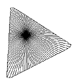
Figure 3: Reconstructed perimeter of triangle
References
(1) Woo et al., J. Biomech Eng, 1988
(2) Thay Lee, MS Thesis, University of California, San Diego, 1986