The Michael Group
University of Pittsburgh

Home                 Research                 Group Members                 Publications                 Contact                 Useful Links

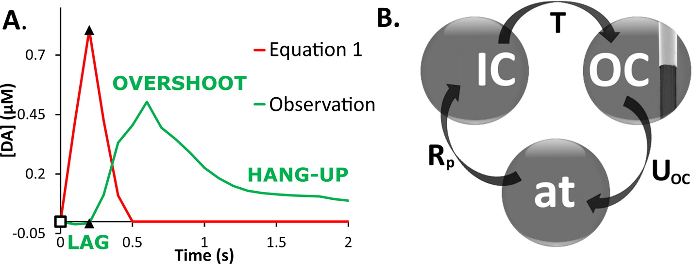
A Novel Restricted Diffusion Model of Evoked Dopamine

In vivo fast-scan cyclic voltammetry provides high-fidelity recordings of electrically evoked dopamine release in the rat striatum. The evoked responses are suitable targets for numerical modeling because the frequency and duration of the stimulus are exactly known. Responses recorded in the dorsal and ventral striatum of the rat do not bear out the predictions of a numerical model that assumes the presence of a diffusion gap interposed between the recording electrode and nearby dopamine terminals. Recent findings, however, suggest that dopamine may be subject to restricted 
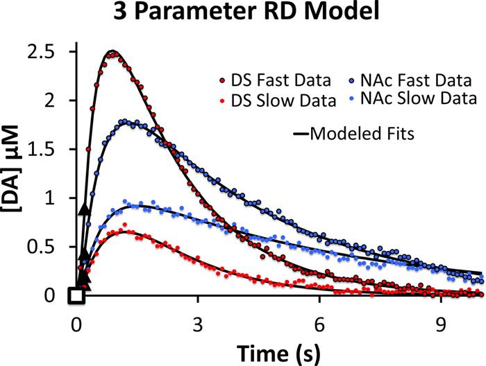
diffusion processes in brain extracellular space. A numerical model cast to account for restricted diffusion produces excellent agreement between simulated and observed responses recorded under a broad range of anatomical, stimulus, and pharmacological conditions. The numerical model requires four, and in some cases only three, adjustable parameters and produces meaningful kinetic parameter values.


                     1
Figure 1: (A) Evoked responses, as predicted by eq 1 (red line), rise during the stimulus and decay back to zero after the stimulus ends. However, observed responses (green line) also exhibit lag (an initial delay in the appearance of the signal), overshoot (the signal continues to rise after the stimulus ends), and hang-up (the signal remains elevated for prolonged periods after the stimulus ends instead of returning to baseline). The open square indicates the start of the stimulus, and the closed triangles indicate the end of the stimulus. (B) Schematic representation of the RD model (see the Methods section for definitions of the parameters). The extracellular space is divided into inner (IC) and outer (OC) compartments. DA is released from axon terminals (at) to the IC, is subsequently transported to the OC, and is removed from the OC by uptake. The model postulates that FSCV recording takes place in the OC.


                                                  2
Figure 2: Three-parameter RD simulations of postnomifensine averaged responses to 0.2 s, 60 Hz stimuli recorded in the dorsal striatum and the nucleus accumbens.