The Michael Group
University of Pittsburgh

Home                 Research                 Group Members                 Publications                 Contact                 Useful Links

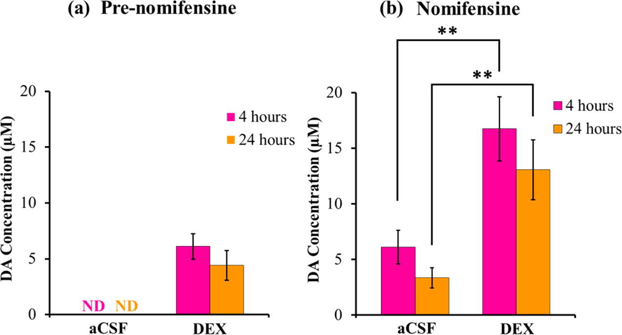
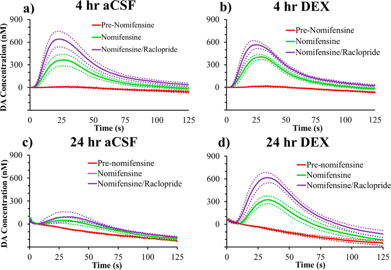
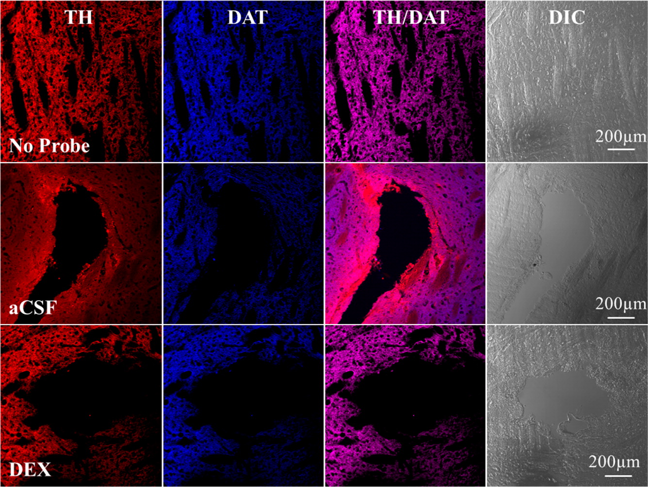
Microdialysis in the Rat Striatum: Effects of 24 h Dexamethasone
Retrodialysis on Evoked Dopamine Release and Penetration Injury


The power of microdialysis for in vivo neurochemical monitoring is a result of intense efforts to enhance microdialysis procedures, the probes themselves, and the analytical systems used for the analysis of dialysate samples. Our goal is to refine microdialysis further by focusing attention on what happens when the probes are implanted into brain tissue. It is broadly acknowledged that some tissue damage occurs, such that the tissue nearest the probes is disrupted from its normal state. We hypothesize that mitigating such disruption would refine microdialysis. Herein, we show that the addition of dexamethasone, an anti-inflammatory drug, to the perfusion fluid protects evoked dopamine responses as measured by fast-scan cyclic voltammetry next to the probes after 24 h. We also show that dexamethasone stabilizes evoked dopamine responses measured at the probe outlet over a 4–24 h postimplantation interval. The effects of dexamethasone are attributable to its anti-inflammatory actions, as dexamethasone had no significant effect on two histochemical markers for dopamine terminals, tyrosine hydroxylase and the dopamine transporter. Using histochemical assays, we confirmed that the actions of dexamethasone are tightly confined to the immediate, local vicinity of the probe.


                       1       k         

Figure 1:  Summary of the amplitudes of evoked DA responses at the E1 location (average +/- SEM) observed 4 h (pink) and 24 h (orange) after probe implantation (a) before and (b) after administration of nomifensine. In the absence of nomifensine, DA was nondetectable (ND) near probes perfused with aCSF, so statistical analysis was confined to the results obtained after nomifensine administration (panel b). Statistical analysis was by two-way ANOVA with time (4, 24 h) and perfusion condition (aCSF, DEX) as factors. Time is not a significant factor (F(1,20) = 2.22, p > 0.05). Perfusion condition is a significant factor (F(1,20) = 22.1, p < 0.0005). The interaction between factors was not significant (F(1,20) = 0.046, p > 0.05). Post hoc pairwise comparisons with Bonferroni correction show that DEX significantly increased the post-nomifensine responses at 4 and 24 h compared to those observed with aCSF (**p < 0.005).

  
                       2                        e

Figure 2: Effect of aCSF and DEX on the average (+/- SEM) evoked DA release measured at the probe outlet pre-nomifensine (red), after nomifensine (green), and after both nomifensine and raclopride (purple). Evoked release was measured (a, b) 4 h and (c, d) 24 h after probe implantation (n = 6 per group).

                                                     3
Figure 3: Separate rows illustrate representative fluorescent images of striatal tissue with no probe, after retrodialysis of aCSF, or DEX for 24 h. Separate columns provide tissue (from left to right) labeled with TH, DAT, their respective overlaid images, and corresponding DIC images. Scale bars are 200 μm.